A. Kumar et al. / Journal of Alloys and Compounds 609 (2014) 100–106
101
yellow emission intensity ratio (B/Y) via selecting different hosts or
changing the doping concentrations of Dy3+ions [9,10].
intensity (observed XRD intensities) and stimulated intensity (cal-
culated XRD intensities) from the pattern shown in Fig. 1a. The
Rietveld refinement is performed for the synthesized NaAlSiO4 dif-
fraction pattern using the P63 structure model reported by Kahlen-
berg et al. [13] which converged to Rp = 14.9%, and Rwp = 19.9%
Therefore, the actual purpose of this work is to explore a sol–gel
combustion synthetic route for the synthesis of single phase NaAl-
SiO4 nanocrystalline host by incorporating Dy3+ ions which is
environmentally safe and less expensive rare earth ion. The crystal
structure of the pure NaAlSiO4, and transformation of phases, mor-
phology and luminescence properties of Dy3+-doped NaAlSiO4
phosphors at various calcinations have been investigated in detail.
It is interesting to study the luminescence of Dy3+ in the NaAlSiO4
host and the energy transfer process between Dy3+and the lumi-
nescent centre present in the NaAlSiO4.
Rexp = 9.67% and goodness of fit (v
2) = 2.1. The experimental, calcu-
lated, and difference XRD patterns are shown in Fig. 1a. NaAlSiO4
crystallizes as a hexagonal structure, (the packing diagram of com-
plete structure presented in the inset of Fig. 1) with a space group
P63 (173) and lattice constants of a = b = 9.97 (3) Å, c = 8.33 (2) Å,
V = 718.23 Å3, Z = 1. Hexagonal nepheline NaAlSiO4 belong to the
group of tecto-silicates, comprises -ABAB- stacking of (Al, SiO4)
sheets [3]. NaAlSiO4 forms three dimensional structure consisting
the framework of AlO4 and SiO4 tetrahedra with each oxygen link-
ing another in a neighboring tetrahedron [14,11] as shown in
Fig. 1c. A schematic of the crystal structure for the NaAlSiO4 host
system along with the strongest reflection observed in the XRD
pattern coming from (201) plane (observed at 2h = 23.181) is also
presented in Fig. 1b. The crystallographic details procured from the
Rietveld refinement of the host NaAlSiO4 are presented in Table 1.
During the process of refinement, the occupancy parameters of the
atoms (Na, Al, Si, and O) comprising the host were evaluated with
reference to their nominal stoichiometric composition. Table 2 pre-
sents the structural parameters of NaAlSiO4.
Fig. 2a shows the XRD diffraction patterns of NaAlSiO4 doped
with 0.5, 1, 1.5 and 2 mol% Dy3+ ion. All the diffraction peaks of
synthesized samples can be assigned to pure hexagonal phase of
NaAlSiO4. The uniform diffraction patterns show that the incorpo-
ration of the Dy3+activators into the host NaAlSiO4 did not affect
the crystal structure of the synthesized material significantly, indi-
cating Dy3+ ions have dissolved completely in the NaAlSiO4 host.
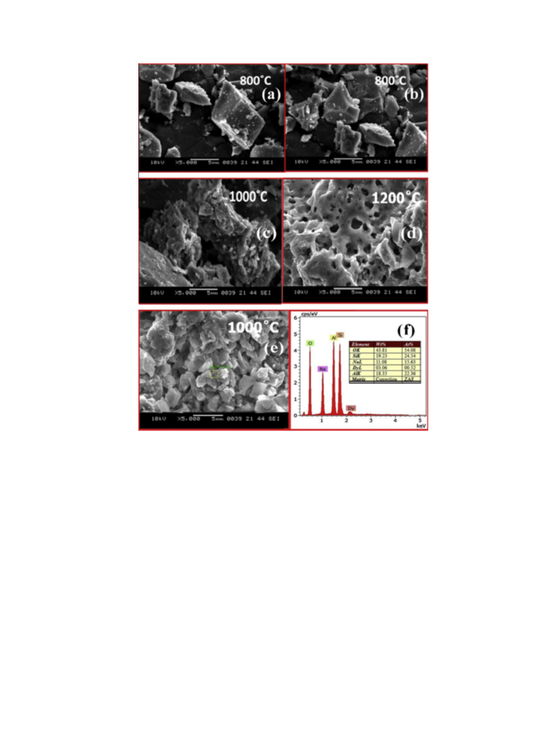
The powder XRD patterns of the pure NaAlSiO4 samples calcined
at different temperatures are shown in Fig. 2b. All the peaks in
the five profiles of the pure samples can be well-indexed to a pure
hexagonal phase of NaAlSiO4. No secondary phase was detected for
the pure samples. The lattice constants for the NaAlSiO4 prepared
at 800 °C are a = 9.97 Å, b = 9.97 Å, and c = 8.33 Å, which is consis-
tent with the International Committee for Diffraction Data (ICDD)
No. 035-0424. It is confirmed that the NaAlSiO4 nanoparticles can
be obtained at as low as 800 °C. It can be noticed that the intensity
of the diffraction patterns becomes sharper at elevated tempera-
tures, indicating increase in the crystallinity of NaAlSiO4 with the
increase of the calcination temperature. All diffraction peaks of
the NaAlSiO4 samples are getting narrowed with temperature
2. Experimental section
2.1. Sample preparation
NaAlSiO4:Dy3+ (Dy = 0.1, 0.5, 1.5, 2 mol%) phosphors were synthesized using
sol–gel combustion synthesis route. The starting materials used for the
synthesis were sodium nitrate (NaNO3 P 99.99%), aluminum nitrate nonahydrate
(Al (NO3)3ꢀ9H2O P 98%), tetraethyl orthosilicate (Si (OC2H5)4 (TEOS, 99.999%),
CH3CH2OH (pure ethanol, P99.8%), dysprosium oxide (Dy2O3). Citric acid was used
as both complexing agent for the gel formation and fuel for the combustion process.
The raw materials used for the synthesis of the phosphors were of analytical grade
and used as received from the commercial sources without further purification.
Tetraethyl-orthosilicate (TEOS) was used as Si source according to the following
reaction equation; Si (OC2H5)4 + 4H2O ? Si (OH)4 + 4C2H5OH in ethanol. First, metal
nitrates of sodium and aluminium were dissolved in distilled water in stoichiome-
tric ratio as per the need of the composition and mixed homogeneously. Second,
TEOS dissolved in ethanol was added to the nitrate solution and they were mixed
together thoroughly. High purity Dy2O3 was dissolved in conc. HNO3 to obtain Dy
(NO3)3 and the solution was evaporated for the removal of the excess of nitric acid,
finally Dy (NO3)3 was added to the mixed solution and heated at 60 °C using a
magnetic agitator under continuous stirring for several hours until it transformed
into transparent sticky gel. The gel residues were then introduced into crucibles
and directly transferred into
a muffle furnace preheated to 600 °C. Due to
occurrence of rapid ignition, the reaction went on vigorously for few seconds with
the evolution of large volume of gases resulting in white fluffy products after the
combustion reaction. The final products were ground to powder in a mortar for
various characterizations. The powders were calcined at temperatures between
800 and 1200 °C for 3 h in ambient air to observe the formation of multiple phases
present in the NaAlSiO4 host lattice.
2.2. Characterization: Structural, optical and morphological properties
The phase purity and the crystallinity of the synthesized samples were obtained
by PANalytical X’Pert Pro diffractometer with Cu Ka radiation (k = 1.541 Å) at 40 kV
and 40 mA over the angular range 10° 6 2h 6 80° at room temperature. Figures of
crystal structure in this work were drawn with VESTA [11]. The photoluminescence
(PL) excitation and emission spectrum were recorded with a Shimadzu Spectroflu-
orometer equipped with Xe lamp as an excitation source at room temperature. The
UV–visible absorption of the phosphors was recorded on Shimadzu 2450 UV–VIS
spectrophotometer equipped with integrating sphere accessory. The morphological
studies were carried out with Scanning Electron Microscopy (SEM), JEOL-6380A and
the elemental composition was determined by using energy dispersive X-ray
spectroscopy (EDS). Transmission electron microscopy (TEM) was observed at an
accelerating voltage of 200 keV on a FEI Tecnai G2 S-Twin. The infrared spectros-
copy of the as prepared was examined using
(spectrum 1000) with KBr pellet.
a Perkin–Elmer spectrometer
3. Results and discussion
3.1. Structural characterization and XRD analysis
High-quality XRD pattern for Rietveld refinement was collected
over an angular 2h range from 10° to 80° at an interval of 0.02°
step. Structural refinement of XRD pattern of NaAlSiO4 is
performed using the Fullprof program [12]. During refinement pro-
cedure, the pseudo-voigt function has been considered in order to
fit several parameters to the data points: such as scale factor, zero
shifting, background, three cell parameters, shape and width of the
peaks, one thermal factor, and two asymmetric factors. A good
agreement was observed between the experimental relative
Fig. 1a. Rietveld analysis of 800 °C calcined NaAlSiO4 nanophosphors.

 Kumar, Ashwini