BioactiVe Metabolites from the Sponge Aka coralliphagum
HR(-)ESIMS t
) 22.9 min, m/z 355.1908 [M - H]- (calcd for
Journal of Natural Products, 2007, Vol. 70, No. 4 509
R
the suspended cells (50 000/mL) were added to 60 µL of serial dilutions
C
22
H
27
O
4
, m/z 355.1904, ∆m ) 1.1).
Siphonodictyal B1 (6): orange powder; [R]D -60 (c 0.501,
MeOH); UV (DAD) λmax 262, 303 (s), 440 nm; IR (KBr) νmax 1648,
of the test compounds. After 5 days the growth was determined using
2
0
16
the MTT assay.
2
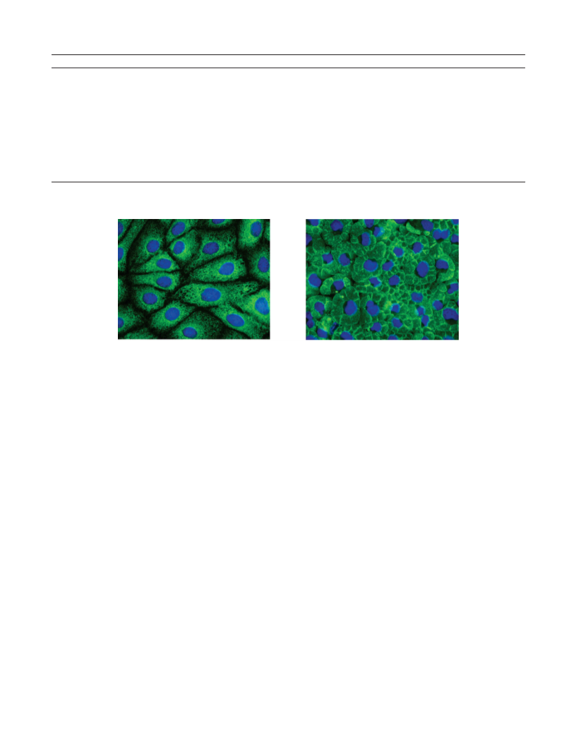
Cell Staining. PtK cells (ATCC CCL-56) grown on glass coverslips
-
1
1
13
1
1
224, 1050 cm ; H NMR data, see Table 2; C NMR data, see Table
were fixed with cold (-20 °C) MeOH/acetone (1:1) for 10 min,
incubated with a primary antibody against GRP-94 (1:1000; Affinity
Bioreagents) and then with a secondary Alexa Fluor 488 goat anti-rat
IgG antibody (1 µg/mL; Molecular Probes), and mounted in ProLong
Antifade Gold (Molecular Probes), which included DAPI to stain the
nuclei.
-
; HPLC/HR(-)ESIMS t
for C24H34NO S
9 2
R
) 12.4 min, m/z 544.1680 [M - H] (calcd
, m/z 544.1669, ∆m ) 2.0).
2
0
Siphonodictyal B2 (7): yellow powder; [R] -76 (c 0.428,
D
MeOH); UV (DAD) λmax 240, 370 nm; IR (KBr) νmax 1647, 1228, 1052
-
1
1
13
cm ; H NMR data, see Table 2; C NMR data, see Table 1; HPLC/
HR(-)ESIMS t
S, m/z 437.1629, ∆m ) 1.7).
Siphonodictyal B3 (8): yellow powder; UV (DAD) λmax 238, 357
R
) 17.1 min, m/z 437.1621 [M - H]- (calcd for
Acknowledgment. We would like to thank B. Hinkelmann (HZI,
Braunschweig) for performing the bioassays, C. Timm (Universit a¨ t
Frankfurt) for measuring optical rotations, R. W. M. van Soest
(University of Amsterdam) for sponge identification, and K. Seifert
22 29 7
C H O
1
13
nm; H NMR data, see Table 2; C NMR data, see Table 1; HPLC/
2
-
HR(-)ESIMS t
, m/z 258.0556, ∆m ) 1.2).
Corallidictyal C/D (9/10): yellow powder; UV (DAD) λmax 220,
R
) 11.9 min, m/z 258.0560 [M - 2H] (calcd for
(
Universit a¨ t Bayreuth) for providing synthetic precursors of siphonod-
22 28 10 2
C H O S
ictyal B.
1
13
2
80, 410 nm; H NMR data, see Table 2; C NMR data, see Table 1;
1
Supporting Information Available: 2D NMR data, 1D H and
-
HPLC/HR(-)ESIMS t
for C22 , m/z 357.2060, ∆m ) 1.9).
Siphonodictyal G (11): light yellow powder; UV (DAD) λmax 220,
R
) 29.6 min, m/z 357.2053 [M - H] (calcd
13
1
13
C NMR spectra of siphonodictyals B1 (6) and B2 (7), H, C HMBC
29 4
H O
spectrum of siphonodictyal B1 (6), MS/MS spectra of siphonodictyals
B1 (6) and B2 (7), and increment calculations. This material is available
free of charge via the Internet at http://pubs.acs.org.
1
13
2
68, 314 nm; H NMR data, see Table 2; C NMR data, see Table 1;
-
HPLC/HR(-)ESIMS t
for C22 S, m/z 421.1679, ∆m ) 3.3).
R
) 19.4 min, m/z 421.1666 [M - H] (calcd
29 6
H O
References and Notes
Hydrolysis of Siphonodictyal B1 (6). A 0.6 mg sample of
siphonodictyal B1 (6) was dissolved in 100 µL of acetic acid (10%)
and incubated for 1 h at RT. Then 5 µL of the test solution and a
(
(
1) R u¨ tzler, K. Smithsonian Contrib. Zool. 1971, 77, 1-37.
2) (a) Sullivan, B.; Djura, P.; McIntyre, D. E.; Faulkner, D. J.
Tetrahedron 1981, 37, 979-982. (b) Sullivan, B. W.; Faulkner, D.
J.; Matsumoto, G. K.; He, C. H.; Clardy, J. J. Org. Chem. 1986, 51,
taurine standard (1 mg/mL in H
gel 60, Merck). The plate was developed with butan-1-ol/acetic acid/
O (80/20/20) as mobile phase. Detection was performed using the
ninhydrin reagent. The R for taurine was 0.26.
2
O) were coated on a Si TLC plate (Si
4
568-4573. (c) Mukku, V. J. R. V.; Edrada, R. A.; Schmitz, F. J.;
Shanks, M. K.; Chaudhuri, B.; Fabbro, D. J. Nat. Prod. 2003, 66,
H
2
f
686-689.
R,R-Diphenyl-â-picrylhydrazyl (DPPH) Assay. The assay was
performed using a modification of a previously described method.14
For UV spectroscopic measurements half microcells were used. To 1000
µL of each sample at different concentrations (200 and 40 µM) in EtOH
were added 250 µL of DPPH (1 mM) in EtOH and 750 µL of EtOH.
The resultant mixture was briefly shaken and maintained at RT in the
dark for 30 min. At the end of this time, the absorbance of the mixture
was measured at 517 nm, using a UV spectrometer. The calculations
(3) Metzger, K.; Rehberger, P. A.; Erben, G.; Lehmann, W. D. Anal.
Chem. 1995, 67, 4178-4183.
(4) Chan, J. A.; Freyer, A. J.; Carte, B. K.; Hemling, M. E.; Hofmann,
G. A.; Mattern, M. R.; Mentzer, M. A.; Westley, J. W. J. Nat. Prod.
1
994, 57, 1543-1548.
(
5) Pretsch, E.; B u¨ hlmann, P.; Affolter, C.; Badertscher, M. Spektrosko-
pische Daten zur Strukturaufkl a¨ rung organischer Verbindungen;
Springer: Berlin, 2001.
(
6) Ragan, M. A. Can. J. Chem. 1978, 56, 2681-2685.
1
4
were performed similarly to literature methods.
(7) Kazlauskas, R.; Murphy, P. T.; Warren, R. G.; Wells, R. J.; Blount,
J. F. Aust. J. Chem. 1978, 31, 2685-2697.
Antimicrobial Assay. Antimicrobial activities were determined by
agar diffusion tests using paper disks of 6 mm diameter soaked with
(8) Urban, S.; Capon, R. J. Aust. J. Chem. 1996, 49, 611-615.
(
9) Dastlik, K. A.; Ghisalberti, E. L.; Skelton, B. W.; White, A. H. Aust.
20 µL of the test compound in MeOH (1 mg/mL). The microorganisms
J. Chem. 1991, 44, 123-127.
were obtained from the HZI collection, grown on standard media, and
cultured in liquid agar medium to a final OD of 0.01 (bacteria) or 0.1
(
(
10) Bernet, A.; Seifert, K. HelV. Chim. Acta 2006, 89, 784-796.
11) Bassett, S.; Ovenden, S. P. B.; Gable, R. W.; Capon, R. J. Aust. J.
Chem. 1997, 50, 1137-1143.
(yeasts). Spores of fungi were collected from well-grown Petri dishes,
which were rinsed with 10 mL of sterile H O. One milliliter of the
2
(
12) Djura, P.; Stierle, D. B.; Sullivan, B.; Faulkner, D. J.; Arnold, E.;
spore suspension was added to 100 mL of molten agar medium. Plates
were incubated at 30 °C, and the diameters of resulting inhibition zones
were measured after 1 and 2 days.
Cell Proliferation Assay. L929 mouse fibroblasts were obtained
from the Deutsche Sammlung von Mikroorganismen und Zellkulturen
Clardy, J. J. Org. Chem. 1980, 45, 1435-1441.
(13) Kang, H. S.; Chung, H. Y.; Jung, J. H.; Son, B. W.; Choi, J. S. Chem.
Pharm. Bull. 2003, 51, 1012-1014.
(14) Fisch, K. M.; B o¨ hm, V.; Wright, A. D.; K o¨ nig, G. M. J. Nat. Prod.
2
003, 66, 968-975.
(
15) Monti, M. C.; Casapullo, A.; Santomauro, C.; D’Auria, M. V.; Riccio,
R.; Gomez-Paloma, L. ChemBioChem 2006, 7, 971-980.
16) Mosmann, T. J. Immunol. Methods 1983, 65, 55-63.
2
(DSMZ) and cultivated at 37 °C and 10% CO in DME medium (high
glucose) supplemented with 10% fetal calf serum. Cell culture reagents
were purchased from Life Technologies Inc. (GIBCO BRL). Growth
inhibition was measured in microtiter plates. Aliquots of 120 µL of
(
NP0603018

 Grube, Achim