
Natural Product Research p. 1598 - 1604 (2016)
Update date:2022-08-16
Topics:
 Tran, Thu Huong
Le Huyen, Tram
Tran, Thi Minh
Nguyen, Tuan Anh
Pham, Thanh Binh
Nguyen Tien, Dat
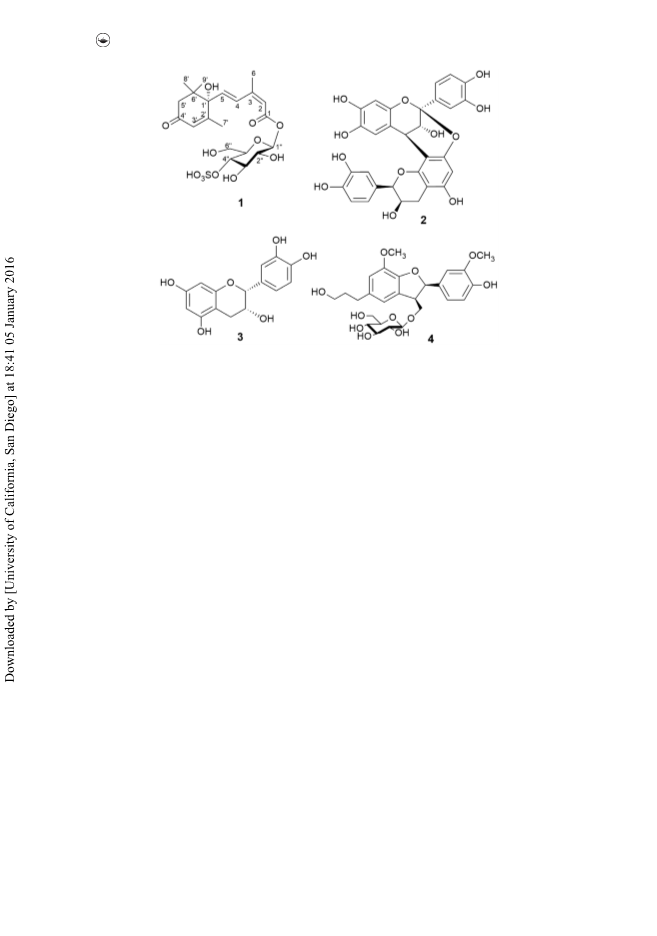
A megastigmane sulphoglycoside together with three phenolic compounds were isolated from the water-soluble fraction of the pericarps of Garcinia mangostana. The structure of the new compound was determined as 4-O-sulpho-β-d-glucopyranosyl abscisate (1) by spectroscopic data. Proanthocyanidin A2 (2) showed potent α-glucosidase inhibitory and DPPH scavenging activities with IC50values of 3.46 and 11.6 μM, respectively.
View More


ClickChem Technology Co., Limited
Contact:+86-0310-6519966/0531-52893837
Address:No.750 Shunhua Road, High-Tech Zone, Jinan city, Shandong China
Frapp's Chemical (NFTZ) Co.,Ltd
Contact:+86-576-86137892
Address:General Chamber of Commercial Building, 159 Wanchang Middle Road, Wenling, Zhejiang, China
VanderArk International Limited
Contact:86-10-82437576
Address:Qing He
shandong zhongke taidou chemical co.,ltd
Contact:86-531-88682301
Address:Jinan shandong Province CHina
Shanghai Dianyang Industry Co.,ltd
Contact:+86 21 6492 4669
Address:Chejing RD, Songjiang District, Shanghai, China
Doi:10.1016/S0040-4020(01)87304-X
(1986)Doi:10.1021/jacs.0c06866
(2020)Doi:10.1021/ja00818a073
(1974)Doi:10.1021/jp302600a
(2012)Doi:10.3390/molecules24122293
(2019)Doi:10.1021/acs.organomet.6b00935
(2017)
