B. Liu et al. / Journal of Solid State Chemistry 230 (2015) 90–94
91
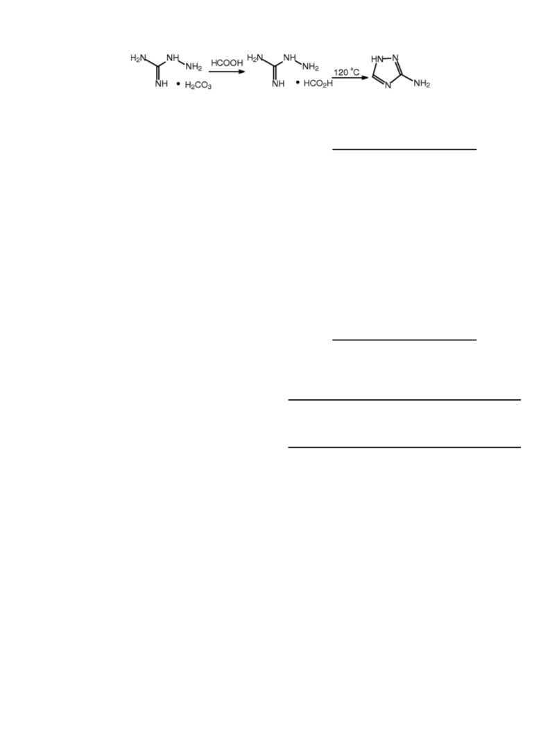
Scheme 1. The synthesis route of 3-amino-1 H-1,2,4-triazole.
Max–3c (Japan) diffractometer (Cu-Kα1,2 X-radiation, λ1
¼
1.540598 Å and λ2¼1.544426 Å), equipped with an X’Celerator
detector and a flatplate sample holder in a Bragg–Brentano para-
focusing optics configuration (40 kV, 40 mA). Intensity data were
collected by the step counting method (step being 0.02°), in con-
Table 1
Crystal and structure refinement data for com-
pound 1.
Empirical formula
C3H9CuN5O1.5
Blue prism
0.24 ꢂ 0.20 ꢂ 0.20
hexagonal
P3221
11.9286(6)
11.9286(6)
10.4690(11)
1290.07(16)
6
202.69
1.565
2.496
618
3.42 to 25.01
8415
1503 (Rint¼0.0229)
Color and Habit
Crystal Size (mm3)
tinuous mode, in the range of 3r2θr60°. Variable-temperature,
Crystal system
solid-state direct current (DC) magnetic susceptibility data down
to 2 K were collected on a Quantum Design PPMS60000 magnet-
ometer. Diamagnetic corrections were applied to the observed
paramagnetic susceptibilities using Pascal’s constants.
Space group
a (Ǻ)
b (Ǻ)
c (Ǻ)
V (Ǻ3)
Z
2.2. Synthesis of 3-amino-1 H-1,2,4-triazole
Fw
Dcalcd (Mgmꢀ3
)
μ (mmꢀ1
F (000)
θ (°)
)
3-Amino-1 H-1,2,4-triazole can be synthesized through con-
densation reaction of aminoguanidine bicarbonate and mathane
acid [13] (Scheme 1) or decarboxylation [14,15]. M. P.¼156–159 °C.
C2H4N4 elemental analysis (%), Found (calcd): C, 28.59 (28.47); H,
4.78 (4.55); N, 66.65 (66.81). FT-IR data (in KBr, cmꢀ1): 3412(w),
3333(w), 3210(w), 3053(w), 2930(w), 2773(w), 2717(w), 1639(s),
1589(m), 1533(s), 1421(m), 1365(w), 1267(m), 1205(m), 1043(s),
965(w), 869(w), 828(w), 724(w), 637(w).
Reflections measured
Independent reflections
Observed Reflection [I42s(I)] 1473
Final R1, wR2 indices (obs.)
R1, wR2 indices (all)
S
0.0577,0.1529
0.0583, 0.1540
1.132
0.017,0.000
1.024, ꢀ0.524
(Δ/s)max/min
(Δρ)max/min (eǺ-3
)
2.3. Synthesis of [Cu(atr)(OH)]·0.5H2O·0.5en (1)
R1¼(Σ||Fo|-|Fc || / Σ |Fo|). wR2¼[Σ (w(Fo2-Fc2)2) / Σ (w
|Fo2|2)]1/2
pH of Hatr ethanol solution (0.085 g, 1 mmol) was adjusted to
8 by ethylenediamine. Aqueous solution containing CuSO4 ꢁ 5H2O
(0.25 g, 1 mmol) was covered with the Hatr solution in a tube.
Over a period of approximate 20 d, the blue crystals were obtained
in the yield of 29.7% (0.06 g). Elemental analysis (%), Found (calcd):
C, 17.85 (17.73); H, 4.41 (4.58); N, 34.52 (34.65). FT-IR data (in KBr,
cmꢀ1): 3399 (b, vs), 2975 (m), 2895 (m), 1631 (m), 1555 (m), 1518
(w), 1451 (w), 1382 (w), 1316 (w), 1271 (w), 1090 (s), 1050 (s), 881
(m), 804 (w), 736 (m), 627 (w), 495 (w).
Table 2
Selected bond distances (Å) and bond angles (°) of compound 1.
Cu1-N11A
Cu1-N14
1.9782(19)
1.982(2)
Cu1-N12B
Cu1-O1
1.989(2)
2.009(2)
88.75(8)
89.08(9)
176.93(9)
N11A-Cu1-N14
N11A-Cu1-N12B
N14-Cu1-N12B
176.50(8)
90.90(6)
91.42(8)
N11A-Cu1-O1
N14-Cu1-O1
N12B-Cu1-O1
Symmetry codes: A¼x-yþ1, -yþ1, -zþ1/3; B¼-yþ1, x-yþ1, z-1/3.
2.4. Single crystal X-ray diffraction
Single crystals of compound 1 were manually harvested from
crystallisation vials and mounted on Hampton Research CryoLoops
using FOMBLIN Y perfluoropolyether vacuum oil (LVAC 25/6, pur-
chased from Aldrich) [16] with the help of a Stemi 2000 stereo-
microscope equipped with Carl Zeiss lenses. Data were collected
on a Rigaku Mercury CCD diffractometer equipped with a gra-
1 were both considered as 3-connected nodes and the topological
network of compound 1 was calculated using the ADS program of
the TOPOS 4.0 Professional structure-topological program
packages.
phite-monochromated Mo-K
(2) K. The intensity data were collected by the
α
radiation (
λ
¼0.71073 Å) at 293
3. Results and discussion
ω
scan technique
and were reduced using CrystalClear program [17]. The crystal
structure of compound 1 was solved by direct method using
SHELXTL™ package of crystallographic software [18] and refined
by full-matrix least-squares technique on F2. All non-hydrogen
atoms were refined anisotropically. Hydrogen atoms were located
at geometrically calculated positions to their carrier atoms and
refined with isotropic thermal parameters included in the final
stage of the refinement. A summary of the structural determina-
tion and refinement for compound 1 is listed in Table 1. The se-
lected bond distances and angles are listed in Table 2.
3.1. Structural description of compound 1
Much interest has been focused on helical supramolecular ar-
chitectures with potential important applications in advanced
materials such as optical devices [20]. Though the achiral approach
almost always leads to a racemic mixture [21], chiral crystals can
still occur through self-assembly, which is not surprising even if
simple salts can crystallize in chiral space group [22]. The inherent
chirality of this architecture comes from spatial disposition rather
than the presence of chiral centers, which can have important
applications in a spontaneous splitting of the racemate on crys-
tallization. In this paper, compound 1 features a chiral metal-or-
ganic framework fabricated by left-handed helices based on
foundational repeating neutral [Cu(atr)(OH)] units. Compound 1
2.5. Topology
According to A. F. Wells’ topology definition [19] and the
MOF structural features, Cu(II) atom and atr‒ ligand in compound

 Liu, Bing